The Human Face
Evolutionary Perspective
I studied cranio-facial evolution way back in graduate school so I’m very interested in this topic. The facial skeleton of modern humans has a number of novel features compared to other ancestral species. Several of these relate to a general reduction in the projection of the upper and middle facial regions. Modern humans have short (front-to-back) faces compared to Neanderthals, a group with whom they co-existed. A recent study reviewed potential drivers of human facial evolution and suggested that physiological, mechanical, and even social (!) factors may have interacted to shape the modern human face. Interestingly, variation in dimensions of the midfacial region has been used to investigate possible hybridization between modern humans and Neanderthals.
Despite the advantages that may have come with facial reduction, a possible downside relates to dental development. In modern humans, the wisdom teeth (3rd molars) have the highest rate of impaction, or failure to emerge correctly. One possible explanation for this is that facial shortening reduced the amount of space in the jaw available for teeth to emerge. A recent study reported a relationship between facial shape in humans and the prevalence of third molar impaction. This points to a common theme in evolutionary studies: a change in a certain structure, while having detrimental effects, can still convey a net benefit.
Functional Grouping of Facial Bones
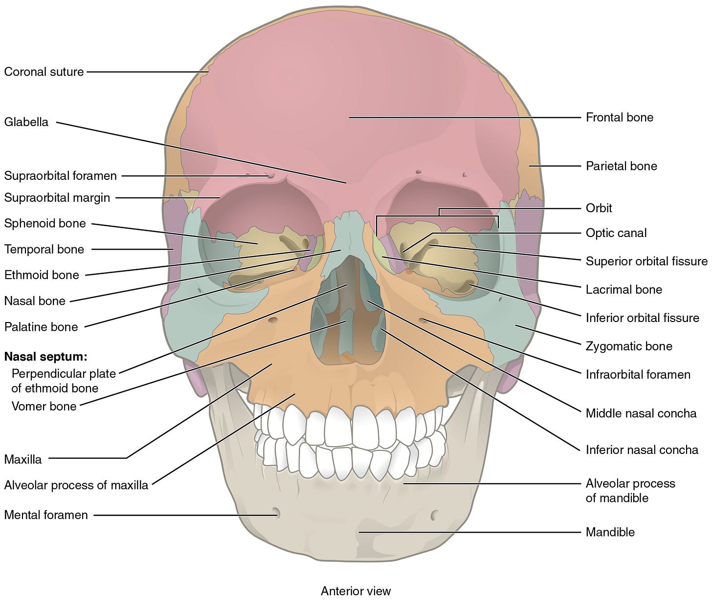
Most of the images linked to (and the one shown below) are from OpenStax (Anatomy and Physiology), an open-source online textbook. All of the bones have separate right and left sides except for the ethmoid, sphenoid, vomer, and mandible.
First, check out this video of a skull with all of the bones separated and labeled.
Bones that Support Chewing
The maxilla (Latin, upper jaw; pleural = maxillae) holds the upper teeth and the mandible (Latin, jawbone) holds the lower teeth. Modern human jaws don’t protrude that much compared to other primates but early human ancestors had large canine teeth which increased how far the maxilla protruded. The zygomatic (Greek, bolt or bar) bone forms the cheekbone and part of the bar, the zygomatic arch, that you can feel on the side of your face. This bone and the arch provide attachment for one of your large chewing muscles. Paranthropus boisei, an extinct member of the human subfamily, had very large back teeth (molars) and large zygomatic arches that allowed for the passage of very large chewing muscles.
Bones that Form the Roof of Your Mouth
The right and left palatine processes of the maxillae come together to form most of the hard palate. The palatine (Latin, palate = vault) bones are the other bones that contribute to the hard palate and they extend the back (posterior) part. Failure of the palatine processes to fuse correctly during development can result in a cleft palate (this results in communication between the nasal and oral cavities). Children born with a cleft palate face challenges related to feeding and speech production and face elevated risk of serious ear infections. However, this defect can be corrected surgically with procedures beginning in early childhood.
Formation of the Eye Socket
A total of 7(!) bones come together to form your eye socket (orbit). These are the: maxilla, zygomatic, frontal, lacrimal, ethmoid, sphenoid, and palatine. The lacrimal (Latin, tear; located in the orbit) bone helps form a canal (the nasolacrimal canal) that drains tears down to the nasal cavity. This is why someone might get a runny nose while crying. The frontal bone is considered part of the neurocranium but does help form part of your orbit and forehead. The ethmoid bone forms part of the inner (toward the middle) wall of the eye socket. This part of the bone can be fractured (blowout fracture) following impact to the face or eyeball.
Spaces Inside Bones
Each maxilla contains a space called the maxillary sinus. Spaces inside certain facial bones, including the maxillae, are called paranasal sinuses. I’ve always been interested in the paranasal sinuses, maybe because of all of the debate regarding their possible function(s). These sinuses all connect to the nasal cavity and are lined with membrane. Thus, the sinuses are susceptible to infections that spread from the upper respiratory tract, potentially resulting in a painful condition called sinusitis. Maxillary sinusitis can even cause tooth pain due to aggravation of nerves near the sinus that supply the upper teeth (have you experienced this?). Other facial bones with sinuses include the ethmoid, sphenoid, and frontal.
Bridging the Face and the Braincase
The sphenoid (Greek, wedge-shaped) bone helps bridge the face and braincase. Go back to the video, you’ll see the sphenoid bone (in yellow) wedged in there. The top surface of the sphenoid bone, in the midline, features a structure called the sella turcica (Latin for ‘Turkish saddle’). The depressed part of the ‘saddle’ is called the pituitary fossa and houses the pituitary gland. In the vast majority of cases, pituitary surgery (e.g., to operate on a pituitary tumor) is performed through a trans-nasal approach. The ethmoid (Greek, sieve; located between the eye sockets) is the other bone that helps bridge the face and braincase. The bottom surface of this bone faces the nasal cavity, the top faces the braincase. The cribriform plate on the top surface of the ethmoid bone has a number of holes that allow passage of olfactory nerves from the nasal cavity into the cranial cavity. I always remembered this structure by thinking of a cribbage board with all of the holes. A traumatic facial injury can cause a fracture in this area, possibly causing rhinorrhea (leakage of cerebrospinal fluid through the nose). A friend of mine suffered this type of injury playing rugby. A fracture of this structure can also damage olfactory nerves and cause anosmia (loss of the sense of smell).
More Details about Facial Bones
Maxilla
Alveolar process - This is the rim of bone where the upper tooth sockets are located. Tooth loss will (eventually) result in extensive remodeling and loss of alveolar bone. Alveoli (singular = alveolus) are the tooth sockets). Alveolus refers to a hollow space; hence, the air sacs in the lungs have the same name.
Infraorbital foramen - This hole [=foramen] is located below the orbit; allows passage of a sensory nerve, the infraorbital nerve). Animals with lots of whiskers have a larger infraorbital nerve!
Ethmoid
Perpendicular plate - Together with the vomer below, this vertical feature forms part of the nasal septum.
Middle and superior nasal concha – The superior nasal conchae are covered with olfactory (sense of smell) mucosa (membrane) where olfactory receptor cells are located. The nasal conchae can be covered with respiratory and olfactory mucosa and the distribution of these varies considerably among species.
Crista galli - This feature allows for attachment of the dura mater, a membrane surrounding the brain. The Latin origin of this name means ‘crest of the rooster’.
Sphenoid
Greater wing – This feature forms part of the side wall of the cranium, in the temple area.
Lesser wing - This structure, totally inside the braincase, helps form the optic canal which allows passage of the optic nerve.
Pterygoid process - This structure is an inferior [downward] projection that bifurcates into medial and lateral pterygoid plates. Pterygoid means ‘shaped like a wing’ (same root as in Pterydactyl).
Body - This is the central part of the sphenoid from which the wings and pterygoid processes project; it contains the sphenoid sinus.
Foramina – The sphenoid bone has several foramina (holes) that allow passage of nerves or blood vessels. These include foramen rotundum (nerve to the face), foramen ovale (nerve to chewing muscles), and foramen spinosum (artery to the dura mater). The superior orbital fissure in the back of the eye socket allows several nerves to pass through.
Mandible
The ramus is the vertical part of the mandible and the body is the horizontal part that houses the lower teeth. The angle is the point where the corpus meets the body.
Mandibular condyle - This is the joint surface that connects to the mandibular fossa of the temporal bone. If you’ve heard of the painful condition called ‘TMJ’, it means temporo-mandibular joint.
Coronoid process - This triangular structure provides attachment for the temporalis muscle (a chewing muscle).
Alveolar process - This is the rim of bone where the lower tooth sockets are located.
Mental foramen - This is a hole that allows passage of a sensory nerve in the chin area.
Mandibular foramen - This hole on the inner surface of the ramus allows passage of a sensory nerve that supplies the lower teeth; an injection near this site can be performed to deaden the nerve (nerve block) for certain dental procedures.
The vomer (Latin, ploughshare = shaped like a plow) is located inside the nasal cavity and forms part of the midline nasal septum that splits the nasal cavity into right and left sides. A septum that is off-center (deviated septum) can impact sleep and cause snoring.
The nasal (Latin, pertaining to the nose) bones form the bridge of the nose. A ‘broken’ nose can involve fractures to these in addition to the nasal cartilage.
The inferior nasal conchae (concha = from Latin [shell]; singular = concha) is located inside the nasal cavity. The middle and superior nasal conchae are part of the ethmoid bone.


